Content:
When you see something you want in the store, you reach for it. When you notice the traffic lights begin to change green, you put your foot down. And while these things seem like completely natural responses to everyday occurrences, there’s a lot more going on in the brain than we give it credit for.
A new study carried out by researchers from MIT’s Picower Institute for Learning and Memory proves how crucial one particular part of the brain is when it comes to transforming seeing into doing. That region is called the posterior parietal cortex (PPC).
As explained by senior author Mriganka Sur, the Paul E. and Lilah Newton Professor of Neuroscience in the Department of Brain and Cognitive Sciences, vision is where it all begins. But then that visual data has to be transferred into motor commands. Sur is hoping that the new study will help to explain why some people who have suffered from stroke or brain injuries experienced a problem called “hemispatial neglect”. This is where people are unable to act upon any objects lying on one side of their field of vision. They can often see the item, their brain just doesn’t recognize the need to do anything.
The study involved using mice to pinpoint exactly how PPC sprang into action. And what they showed was that it contained a mix of neurons that was accustomed to the process visuals, make decisions, and take action. “This makes the PPC an ideal conduit for flexible mapping on sensory signals onto motor actions, says co-lead author of the study, Gerald Pho, former graduate student in the Sur lab who’s now at Harvard University.
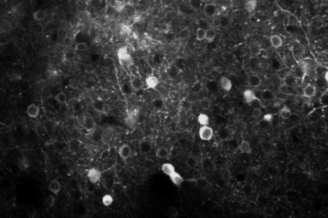
During the trial, the mice were given one simple task to compete and that was if they saw a striped pattern move upward on the screen, they were to lick a nozzle to receive a liquid reward. If the stripes moved sideways, they were to not lick. If they did, they would get a bitter liquid as opposed to the reward. As they carried out the test, researchers recorded the neuron activity of the mice in two areas of the brain: that which processes sight, the visual cortex; and that which receives input from the visual cortex and other regions, the PPC.
The researchers found that in both regions the cells glowed more brightly after becoming active, indicating quite clearly at what stage they got involved. Those in the visual cortex seemed to light up when a pattern emerged and moved, whereas those in the PPC showed more variety in the way they responded. A few of them (around 30 percent) acted in the same way as the visual cortex neurons, becoming active when a pattern moved the right way. But most were randomly responsive. Not just about seeing something, but in the chance to act upon it also.
“Many neurons in the PPC seemed to be active only during particular combinations of visual input and motor action,” explains co-lead author Michael Goard, a former MIT postdoc now at UC Santa Barbara. “This suggests that rather than playing a specified role in sensory or motor processing, they can flexibly link sensory and motor information to help the mouse respond to their environment appropriately.” However, even the odd error was instructive, suggesting that a lot of PPC neurons are in fact oriented towards acting.
Photo: